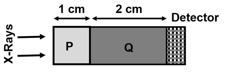
A normally incident X-ray of energy 140 keV passes through the tissue phantom shown. Tissue P has attenuation coefficient 1 cm⁻¹ and thickness 1 cm; tissue Q has coefficient 10 cm⁻¹ and thickness 2 cm. Calculate detected intensity, expressed as energy in μeV under the source’s attenuation model, to one decimal place.
Apply exponential attenuation successively: Eout=140exp[−(1×1+10×2)] keV=140e⁻²¹ keV. One keV equals 10⁹ μeV, so Eout=140×10⁹e⁻²¹≈106.156 μeV, or 106.2 μeV. This source convention attenuates total beam energy/intensity; it does not mean each surviving photon loses energy by this exponential factor.
More X-Ray and Computed Tomography questions
GATE 2023 BM Q57– A two-dimensional square plate of side 20 mm contains a homogeneous circular inclusion of…
GATE 2022 BM Q64– A first-generation CT scanner has a point source and detector translating together at 0.5…
GATE 2022 BM Q65– An acceptable chest radiograph is obtained at source distance 0.75 m using 50 kVp and 50…
GATE 2021 BM Q28– Which statements about planar X-ray imaging are correct?
GATE 2021 BM Q63– An 80 kVp X-ray system uses a 2.5 mm aluminium filter, replaced by copper for equal…
GATE 2020 BM Q16– In diagnostic X-ray imaging, which is NOT a primary electromagnetic-radiation interaction…