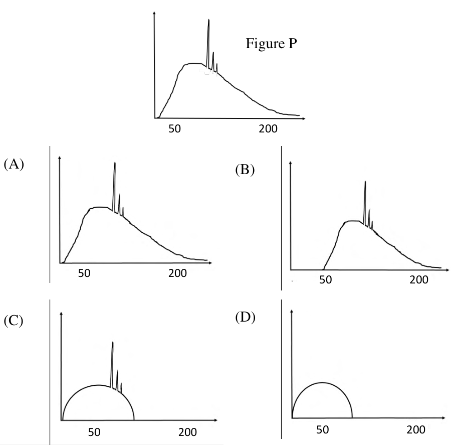
A polychromatic X-ray beam has the energy spectrum in Figure P. Which option depicts the energy spectrum after passing through a human body? In every graph the horizontal axis is energy in keV and the vertical axis is relative X-ray intensity.
Low-energy photons are preferentially absorbed, reducing intensity and increasing the mean energy of the transmitted spectrum: beam hardening. The upper energy endpoint remains fixed by the source voltage. B suppresses the low-energy part while retaining the high-energy extent and characteristic lines. C and D instead truncate the high-energy end.
More X-Ray and Computed Tomography questions
GATE 2023 BM Q40– A parallel X-ray beam traverses a uniform circular disc of radius R and linear attenuation…
GATE 2023 BM Q56– A normally incident X-ray of energy 140 keV passes through the tissue phantom shown.…
GATE 2023 BM Q57– A two-dimensional square plate of side 20 mm contains a homogeneous circular inclusion of…
GATE 2022 BM Q64– A first-generation CT scanner has a point source and detector translating together at 0.5…
GATE 2022 BM Q65– An acceptable chest radiograph is obtained at source distance 0.75 m using 50 kVp and 50…
GATE 2021 BM Q28– Which statements about planar X-ray imaging are correct?