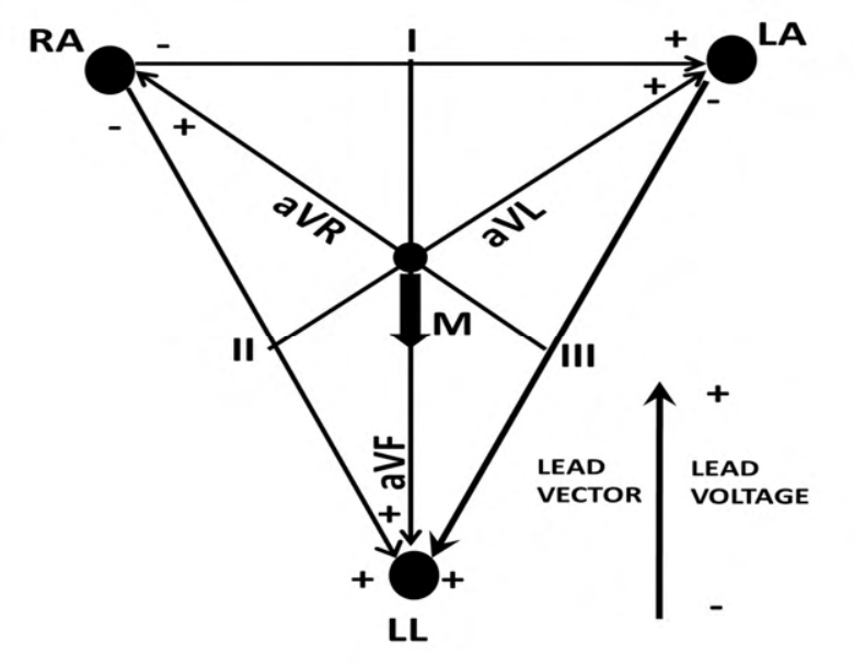
In the shown Einthoven frontal ECG triangle, augmented lead vectors bisect bipolar lead vectors. At the R-wave peak, cardiac vector M has magnitude 5 mV and points vertically downward. Find voltages in leads I and II, respectively, in mV.
Lead I is horizontal, so the downward vector projects to zero on it. Lead II runs from RA to LL, thirty degrees away from downward vertical. Its projection is 5cos30°=4.330 mV with the displayed positive polarity. Thus the ordered pair is (0,4.33), A.
More Biopotentials and Electrodes questions
GATE 2021 BM Q48– Compared with ECG amplifiers, surface-EMG amplifiers have which typical characteristics?
GATE 2020 BM Q22– How many electrodes are used to record a standard 12-lead ECG?
GATE 2020 BM Q36– A mammalian nerve cell initially rests at −80 mV. A drug lowers intracellular K⁺ from 150…
GATE 2020 BM Q65– A cell is driven by current i(t)=u(t). Its linear membrane model has impulse response…
GATE 2026 BM Q43– An EEG PSD has band-average values (μV²/Hz): 0.4–4 Hz:3; 4–8:4; 8–13:8; 13–30:5; 30–100:3.…
GATE 2026 BM Q44– Match ECG waveforms X,Y,Z in Panel 1 with (i) normal condition, (ii) atrial flutter, (iii)…