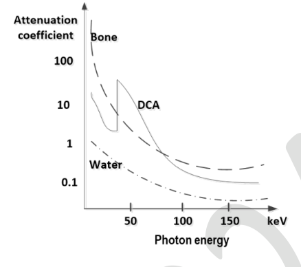
The graph shows X-ray attenuation coefficients versus photon energy for bone,DCA and water. A phantom is imaged at 50 and 150 keV and the developed film viewed. Which statements hold?
Correct answer: (B) At 50 keV DCA appears brighter than bone; (C) At 150 keV bone appears brighter than DCA
Explanation
Greater attenuation exposes the film less, so the region appears brighter on the displayed conventional radiograph. At fifty keV the graph places DCA above bone, making DCA brighter. At one hundred fifty keV bone lies above DCA, reversing the comparison. Thus B,C.
More X-Ray and Computed Tomography questions
GATE 2024 BM Q53– A 70 keV X-ray beam passes through 16 cm muscle and 4 cm bone. Their half-value layers are…
GATE 2023 BM Q23– A polychromatic X-ray beam has the energy spectrum in Figure P. Which option depicts the…
GATE 2023 BM Q40– A parallel X-ray beam traverses a uniform circular disc of radius R and linear attenuation…
GATE 2023 BM Q56– A normally incident X-ray of energy 140 keV passes through the tissue phantom shown.…
GATE 2023 BM Q57– A two-dimensional square plate of side 20 mm contains a homogeneous circular inclusion of…
GATE 2022 BM Q64– A first-generation CT scanner has a point source and detector translating together at 0.5…